What is Breast Imaging?
Breast imaging encompasses non-invasive tests that produce detailed images of the breast’s internal tissues. It is a critical tool for screening, diagnosing, and managing breast conditions, particularly breast cancer. Early detection through imaging often identifies abnormalities before physical symptoms appear, allowing timely intervention and better treatment outcomes.
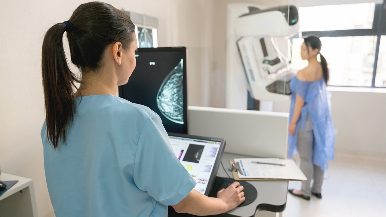
Types of Breast Imaging
1. 3D Mammogram (Digital Breast Tomosynthesis): Captures multiple X-ray images to create a three-dimensional view of the breast. Especially effective for women with dense breast tissue, it improves detection accuracy, reduces false positives, and identifies tumors earlier than traditional 2D mammograms.
2. Breast Ultrasound: Uses sound waves to produce real-time images. It helps distinguish solid masses from fluid-filled cysts, evaluate lumps found in mammograms, and guide biopsies. Safe and radiation-free, it is suitable for all ages.
3. Breast MRI (Magnetic Resonance Imaging): Uses magnetic fields and radio waves to generate detailed images, often with contrast dye. Ideal for high-risk patients or further evaluation of suspicious areas, MRI can detect cancers not visible on mammograms or ultrasounds.
4. Image-Guided Biopsies: Combines imaging with minimally invasive needle biopsy to accurately sample tissue from abnormal areas. It is highly precise and less invasive than surgical biopsy.
5. Diagnostic Mammogram: Focuses on specific areas of concern to provide detailed images for accurate evaluation of lumps, pain, or other changes.
How Breast Imaging Affects the Body
● Physical Impact: Most tests are safe, with mild discomfort during mammograms. Mammograms and some biopsies use low-dose radiation, while ultrasounds and MRIs are radiation-free.
● Emotional Impact: Tests can cause anxiety while awaiting results, but early detection brings reassurance and better treatment options.
● After Effects: Image-guided biopsies may cause mild bruising or swelling, usually resolving quickly. Most imaging tests do not require recovery time.
Diet & Lifestyle Tips
● Before Imaging: Stay hydrated, avoid caffeine to reduce breast tenderness, eat a light meal, skip deodorants or lotions, and wear comfortable two-piece clothing.
● After Imaging: Resume normal diet, increase fluids if contrast dye was used, engage in mild activity, rest if a biopsy was performed, and monitor for side effects.
When to See a Doctor
● New lumps or thickening in the breast or underarm
● Changes in breast size, shape, or appearance
● Persistent pain or tenderness
● Nipple discharge, especially bloody or spontaneous
● Nipple inversion or sudden positional changes
● Skin dimpling, puckering, redness, rash, or swelling
● Persistent itching or warmth around the breast
● Family history of breast cancer and uncertainty about your risk
Preventive Check-ups
Even without symptoms, regular clinical breast exams and imaging—like mammograms—are essential, particularly for women over 40 or those with a family history of breast cancer.
Conclusion
Breast imaging is more than a diagnostic tool—it is a cornerstone of proactive breast health. From routine 3D mammograms to advanced image-guided biopsies, modern imaging techniques allow early detection, accurate diagnosis, and timely intervention. By staying informed, recognizing changes, and following a healthy lifestyle, you can protect your breast health and ensure peace of mind.